PBMC Isolation and Analysis

Peripheral blood mononuclear cells (PBMCs) have the characteristics of concentrating various immune cells such as T cells, B cells, NK cells, monocytes, and dendritic cells while excluding large portions of blood such as RBCs, granulocytes and plasma. They are suitable for evaluation of rare cells such as antigen-specific T cells, and can be stored for long periods by cryopreservation. Due to these characteristics, PBMCs have attracted attention in recent years for various applications, including immunological study and vaccine development.

In general, PBMCs are separated and collected from fresh blood by density-gradient centrifugation. The layering of blood on density solutions, and the collection and washing of separated PBMCs are performed manually, requiring skilled operation techniques to obtain PBMCs of high quality and homogeneity. Furthermore, in order to obtain PBMCs that more closely reflect the in vivo state, it is necessary to have an operational system to carry out the process from blood collection to PBMC isolation in as short a time as possible. These points are extremely important to obtain accurate analytical results from studies using PBMCs.

For many years, we have been entrusted with PBMC isolations in clinical trials. We pick up blood samples from study sites, and highly skilled researchers conduct the PBMC isolation process at our GLP laboratory located in Itabashi, Tokyo. In addition to the standard process, we can utilize other simpler isolation devices such as Vacutainer CPT by BD, Leucosep tube by Greiner, and SepMate by STEMCELL Technologies.

With isolated PBMCs, we can conduct analysis by flow cytometry, ELISpot, and qPCR. In addition, we can conduct long-term storage (-80℃, -150℃, liquid nitrogen) for future studies. Examples of our analytical services using PBMCs are shown below.

Examples of Analytical Services using PBMCs

Multiparameter analysis by cytometry

We offer analytical services of cell surface antigens and intracellular antigens by cytometry.
Flow cytometry (FCM) with fluorescently labeled antibodies supporting multiparameter analysis of up to 8 to 12 colors is available and can be used for each subset analysis, phenotyping of immune cells, and analysis of drug stimulation response.

Since mass cytometry has an advantage of less interaction between reagents by using antibodies labeled with stable metal isotopes, analysis of more than 40 parameters is possible from a single sample. Obtained data can be subjected to multivariate analysis (t-SNE, FlowSOM) using FlowJo analyzing software from BD. Please feel free to consult us for details.

Example of manual analysis of CyTOF data

Thirty-seven parameters are displayed on 2 dimensions by dimension reduction (t-SNE analysis) [FlowJo Ver10, FCM analyzing software]

Cells which have similar antigen expression characteristics form a cluster, and that makes it easy to visualize existing cell groups and grasp complicated high dimension information.
In this case, 2 groups of major T cells and 3 other groups are recognized by a heat map display of CD3 expression.

Analysis of cytokine producing cells by ELISpot

Enzyme-Linked ImmunoSpot (ELISpot) is an immunoassay with very high sensitivity which can detect cytokines secreted from a single cell.
Cytokines secreted from cells are trapped by antibodies on the well bottom layer, then react with secondary antibodies and enzymes, and form cytokine spots at the position of the well bottom layer where the cells are located. Cytokine producing cells can be visualized and determined by counting these spots. As the cytokines secreted from cells combine with antibodies immediately, this technique is free from any influence of degradation or uptake into cells.
This method is useful in research of small numbers of activated cells observed in some specific immune reactions, and we have a lot of experience in efficacy evaluation studies for development of vaccines for infection and cancer, and cell pharmaceuticals.

Immunospot Analyzer

Evaluation of cellular responsiveness by immunoassay

Immune responsiveness is evaluated by immunoassay of cytokines and chemokines secreted from PBMC derived from patients. Stimulation response can be evaluated by analyzing biomarkers in the culture supernatant of isolated PBMC stimulated with drugs or peptides.

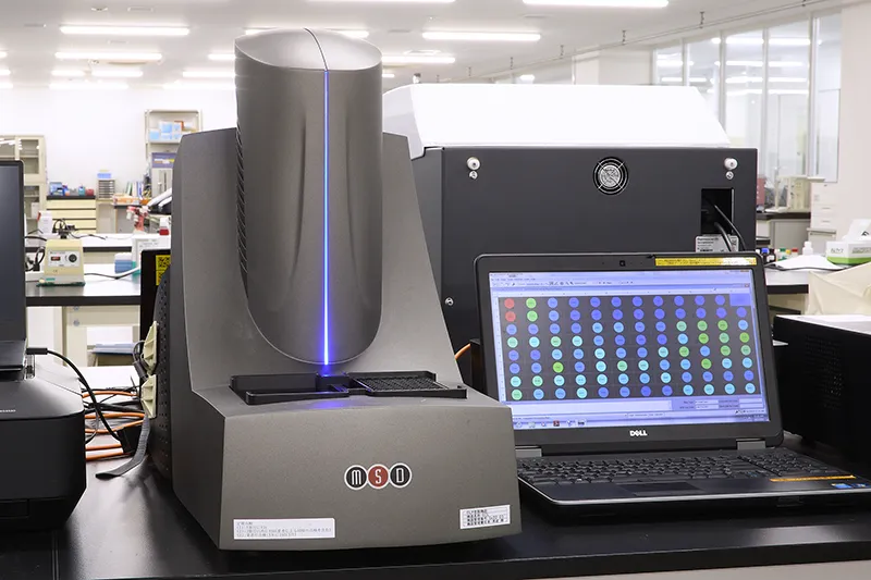
For Biomarker Analysis, in addition to single marker analysis by ordinary ELISA, simultaneous determination of multiple markers by Multiarray System from Meso Scale Diagnostics and Bioplex Suspension Beads Array System from Bio-Rad are also effective.

MESO QuickPlex
MESO QuickPlex
BioPlex
BioPlex