Best Practice hERG Assay

Best Practice hERG Assay

hERG channel plays an important role in cardiac safety assessment. Mediford performs Best Practice hERG assay in compliance with GLP (Good Laboratory Practice) according to the new ICH S7B Guideline Q&As. Our staff’s proficiency with the patch-clamp method provides our clients with high-quality data quickly and efficiently.

With Exceptional Performance, Outstanding Skills, and Meticulous Attention, Mediford Provides Patch-clamp Assay Tailored to Meet Our Clients’ needs.

The patch-clamp method, a technique for measuring cellular electrical activity, has been widely used in a variety of fields such as basic research and drug discovery. Mediford mainly provides study services for evaluating the effects of drugs on cardiac ion channels using cardiac ion channel-expressing cells and the accurately measurable manual patch-clamp platform. We offer evaluations from a wide range of implementation standards, from screening to GLP. Furthermore, we can also conduct studies under customized conditions and develop assay systems according to our clients’ needs.

Mediford always strives to improve the quality of our patch-clamp assay services and to ensure our services are tailored to meet our clients’ needs. Please feel free to contact us.

Click here to contact us

Exceptional performance

To date, mediford has consistently demonstrated exceptional performance in both quality and quantity of GLP hERG assays, earning high ratings from our valued clients. The hERG study, which is particularly important among in vitro cardiovascular studies, is a safety pharmacology core battery study conducted prior to the first-in-human dosing. In Jan. 2024, mediford launched a new study service, Best Practice hERG assay (GLP-compliant). From the perspective of proarrhythmic risk assessment based on the CiPA paradigm, there is a global trend towards integrating multiple ion channels assay—including Na channels (Nav1.5) and Ca channels (Cav1.2)—with in silico proarrhythmic models, moving beyond sole reliance on hERG channel. Thus, ion channel evaluation using the patch-clamp method in safety pharmacology studies is becoming increasingly diverse. Mediford is currently striving to establish Best Practice patch-clamp assays for Nav1.5 and Cav1.2.

Outstanding skills

Our highly-skilled staff performs measurements efficiently and submits flash reports expeditiously, eliminating concerns about the low throughput of the manual patch-clamp method. Taking into account the properties of drugs and target ion channels, we offer optimal study designs based on our accumulated expertise of more than 20 years of services in patch-clamp and cell culture methods.

Meticulous attention

Our greatest strength is the ability to provide the highest level of attention to detail. Our experienced study directors well-versed in the patch-clamp method and knowledgeable about relevant regulatory guidelines are adept at accommodating requests, swiftly addressing unforeseen situations, and ensuring the study meets our clients’ expectations. We specialize in assay development for pharmacological efficacy studies and/or novel studies on target molecules (ion channels) from the ground up, based on the properties of the drugs and ion channels. Please do not hesitate to contact us if you have any questions about patch-clamp assays.

Delivery and study schedule (hERG assay)

hERG screening assay (non-GLP, room temperature)

Standard study volume

3 compounds
Cumulative application of 2 concentrations (5 min/concentration)
N=2/compound (6 cell data in total)

Workflow
Solubility test 1 to 2 days (as necessary)
Preparation of study protocol
Study implementation Measurement 1 day
Flash report Approximately 1 week from study initiation
Draft report Approximately 1 month from study initiation
(limited to summary and Table)

GLP hERG assay (room temperature)

Standard study volume

Vehicle control, test substance (3 concentrations), positive control
N=5/group (25 cell data in total)

Workflow
Solubility test 0.5 days
(conducted within analytical validation study)
Preparation of study protocol
Study implementation Measurement 1 to 2 weeks
Flash report Approximately 1 month from study initiation
(approximately 2 weeks after completion of measurement)
Draft report 2 to 2.5 months from study initiation
(1 to 1.5 months after completion of measurement)
Final report 3 to 3.5 months from study initiation
(2 to 2.5 months after completion of measurement)

GLP hERG assay (Best Practice, near physiological temperature)

Standard study volum

Vehicle control, test substance (3 concentrations), positive control (4 concentrations, cumulative application of 2 concentrations)
N=4/group (24 cell data in total)

Workflow
Solubility test 0.5 days
(conducted within analytical validation study)
Preparation of study protocol
Study implementation Measurement 3 to 4 weeks
Flash report Approximately 1.5 months from study initiation
(approximately 2 weeks after completion of measurement)
Draft report 2.5 to 3 months from study initiation
(1.5 to 2 months after completion of measurement)
Final report 3.5 to 4 months from study initiation
(2.5 to 3 months after completion of measurement)

Glossary

Safety pharmacology core battery studies

Safety pharmacology studies are designed to investigate the potential undesirable pharmacodynamic effects of drugs on physiological functions. The studies to examine cardiovascular, central nervous, and respiratory systems, which are critical to supporting life, are designated as core battery studies and must be conducted under GLP-compliance prior to clinical trials.

Patch-clamp method

Patch-clamp, a laboratory technique in electrophysiology, was developed by Erwin Neher and Bert Sakmann (Nobel laureates in Physiology or Medicine, 1991). This method allows for precise measurement of electrical activity across biological membranes by achieving an extremely high seal resistance of >1 GΩ between a glass pipette electrode filled with buffer solution and the cell membrane to minimize leakage current. The cell can be clamped, keeping the cell membrane potential constant while recording ionic currents (voltage clamp). Alternatively, the cell membrane potential can be recorded while a constant current is being injected into the cell (current clamp). This technique is widely used to investigate the electrical properties of cells in detail.

hERG (human ether-a-go-go-related gene)

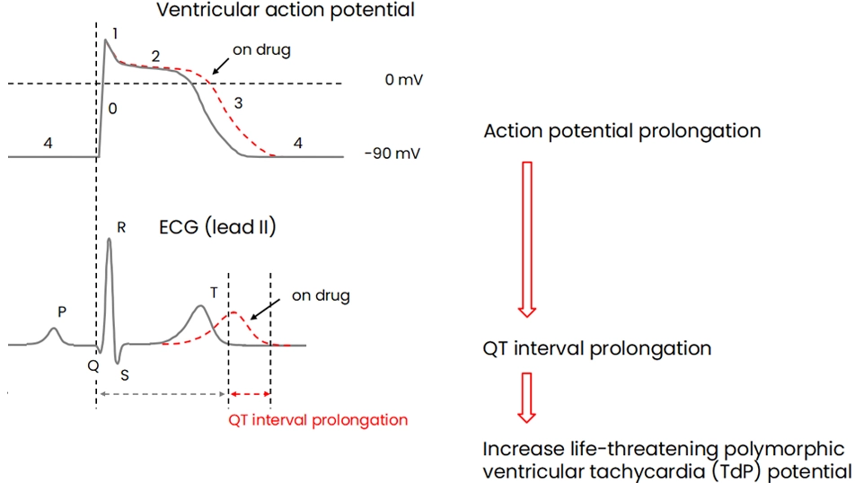
hERG channels are selectively permeable to potassium ions and play an important role in repolarizing the action potential of cardiomyocytes (Figure 1). It is known that QT prolongation due to delayed repolarization of cardiac action potentials can increase the risk of drug-induced lethal polymorphic ventricular tachycardia (TdP) (Figure 2). Therefore, the drug candidates with hERG inhibition often are rejected from further development due to safety concerns, which are regarded as missed opportunities for new drug discovery.

Figure 1:Cardiac action potential and ionic currents

Cardiac action potential and ionic currents
Ionic current Ion channels Physiology
INa Nav1.5 Phase 0 (overshoot)
Ito Kv4.2/4.3 Phase 1
ICa Cav1.2 Phase 2 (plateau), contraction
IKr hERG Phase 3 (repolarization)
IKs KvLQT1/minK Phase 3 (repolarization)
IK1 Kir2.1 Phase 4 (resting potential), relaxation
  • Overshoot: For membrane potential to depolarize beyond 0 mV.
  • Inward current: Ionic currents that flow into the cell and depolarize membrane potential.
  • Outward current: Ionic currents that flow out of the cell and repolarize/hyperpolarize membrane potential.

Figure 2:QT interval prolongation associated with hERG block

QT interval prolongation associated with hERG block
  • QT interval: Time from the start of the Q wave to the end of the T wave. QT interval prolongation is attributed to delayed repolarization (Phase 3) and prolonged plateau (Phase 2).
  • Polymorphic ventricular tachycardiaArrhythmia induced by consecutive ectopic impulses within the ventricles. Torsades de Pointes (TdP) is a form of polymorphic ventricular tachycardia in the setting manifested by QT interval prolongation.

GLP

A set of rules and criteria for a quality system to ensure the reliability when conducting non-clinical safety studies.

ICH S7B Guideline

Describes a non-clinical testing strategy for assessing the potential of a test substance to delay ventricular repolarization. In 2022, the associated Q&As and Training Materials were adopted.

Best Practice

The ICH S7B Guideline Q&As 2.1 provides Best Practice recommendations for patch-clamp assays including those involving hERG channel. To date, each facility has conducted hERG assays using its own methods. However, the adoption of Best Practice is expected to harmonize procedures and reduce data variability across facilities. Non-clinical data recorded under Best Practice will be used for integrated risk assessment alongside clinical findings.

CiPA(Comprehensive in vitro Proarrythmia Assay)

An innovative paradigm for better predicting proarrhythmic risk of drugs in combination with assays of multiple cardiac ion channels assay including hERG channel, simulation of in silico proarrhythmic models using the patch-clamp assay results, and human iPS cell-derived cardiomyocytes. This is based on proarrhythmogenicity and expected to serve as a new paradigm to replace the original guideline strategy (S7B) that focuses on delayed ventricular repolarization, or QT prolongation.

In vitro Cardiovascular Safety Pharmacology Studies

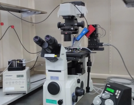
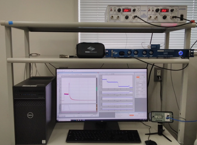
The electrical excitation, or action potential formation, of cardiac myocytes results from the opening and closing of various ion channels (Figure 1). hERG channel plays an especially important role in terminating and repolarizing myocardial action potentials. Significant inhibition of hERG channel by drugs induces broadening myocardial action potential duration and prolongation of QT interval on ECG. This increases the risk of lethal Torsades de Pointes (TdP), which is a major concern in drug development (Figure 2). Patch-clamp assays using HEK293 cells expressing various cardiac ion channels are employed to evaluate the effects of drugs on specific ion channels. The effects of drugs on ion channels are evaluated using either single or cumulative application method. We offer reliable ion channel evaluations using the manual patch-clamp method (Figure 3), the gold standard of patch-clamp methods.

Figure 1:Cardiac action potential and ionic currents

Cardiac action potential and ionic currents
Ionic current Ion channels Physiology
INa Nav1.5 Phase 0 (overshoot)
Ito Kv4.2/4.3 Phase 1
ICa Cav1.2 Phase 2 (plateau), contraction
IKr hERG Phase 3 (repolarization)
IKs KvLQT1/minK Phase 3 (repolarization)
IK1 Kir2.1 Phase 4 (resting potential), relaxation
  • Overshoot: For membrane potential to depolarize beyond 0 mV.
  • Inward current: Ionic currents that flow into the cell and depolarize membrane potential.
  • Outward current: Ionic currents that flow out of the cell and repolarize/hyperpolarize membrane potential.

Figure 2:QT interval prolongation associated with hERG block

QT interval prolongation associated with hERG block
  • QT interval: Time from the start of the Q wave to the end of the T wave. QT interval prolongation is attributed to delayed repolarization (Phase 3) and prolonged plateau (Phase 2).
  • Polymorphic ventricular tachycardiaArrhythmia induced by consecutive ectopic impulses within the ventricles. Torsades de Pointes (TdP) is a form of polymorphic ventricular tachycardia in the setting manifested by QT interval prolongation.

Figure 3:Manual patch-clamp measurement system

Manual patch-clamp measurement system
Inverted microscope equipped with perfusion pump and headstage-attached manipulator
Manual patch-clamp measurement system
PC for data acquisition/analysis, amplifier, and temperature controller
  • Perfusion: Continuous delivery of extracellular fluid to cells immersed in a recording chamber.
  • AmplifierAn electronic circuit or device for amplifying small currents passing through ion channels.

Conventional hERG assay

hERG assay conducted under the original ICH S7B Guideline.

Overview of conventional hERG assay

Item Condition
Platform Manual patch-clamp
Test system hERG-expressing HEK293 cells
Assay grade
  • GLP
  • Screening
Measurement
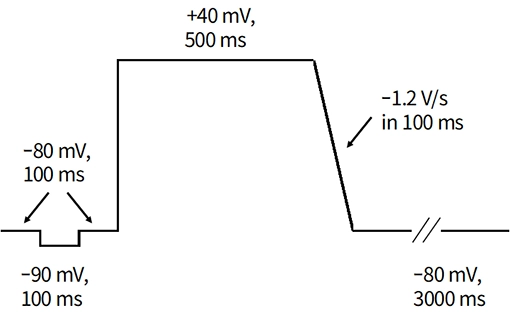
  • Step-pulse voltage protocol
  • at 22-26°C
  • at 0.0167 Hz (every 15 s)
positive control E-4031 (one concentration)
Vehicle
  • DMSO (up to 0.3 v/v%)
  • H2O
Number of data per concentration Minimum number of 5 per test group
Standard assay volume (group) Test article (3 or 4), vehicle control (1), positive control (1); totaling 25 or 30 cells
Data analyses
  • Inhibition rate
  • IC50

Step-pulse voltage protocol

Step-pulse voltage protocol

Best Practice hERG assay

We offer GLP hERG assay services in compliance with the ICH S7B Guideline Q&As 2.1 and related documents (Training Materials and FDA’s Best Practice guidance). Mediford recommends that Best Practice GLP hERG assay be conducted prior to first-in-human studies to prevent repeating GLP hERG assays in the later stage of clinical development if the drugs are expected/intended to have TQT studies waived or alternative TQT studies in animals based on integrated risk assessment.

Review the poster (2024)

Overview of Best Practice hERG assay

Item Condition
Platform Manual patch-clamp
Test system hERG-expressing HEK293 cells
Assay grade
  • GLP
  • Screening
Measurement

FDA’s Best Practice guidance (July 30, 2021)

  • Step and ramp-pulse voltage protocol
  • at 35-37°C
  • at 0.2 Hz (every 5 s)
3 select reference drugs
(positive control underscored)
  • Moxifloxacin
  • Ondansetron
  • Dofetilide (4 concentrations each)
Vehicle
  • DMSO (up to 0.3 v/v%)
  • H2O
Number of data per concentration Minimum number of 4 per test group
Standard assay volume (group) Test article (3 or 4), vehicle control (1), positive control (2); totaling 24 or 28 cells
Data analyses
  • Inhibition rate
  • IC50 and Hill coefficient (95% confidence intervals)
  • Threshold (Pooled safety margin from 3 reference drugs)

Step and ramp-pulse voltage protocol

Step and ramp-pulse voltage protocol

Background data for Best Practice hERG assay

Moxifloxacin

Used as positive control in GLP studies

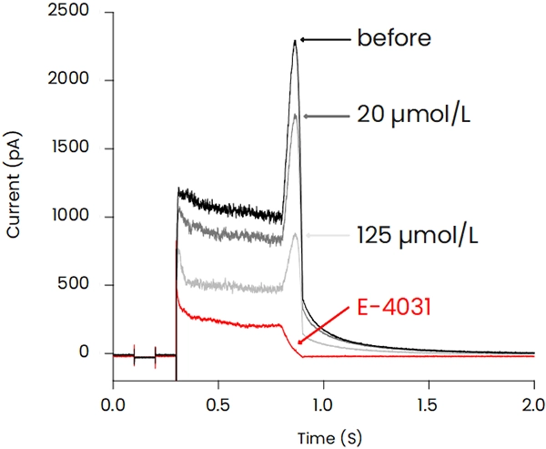
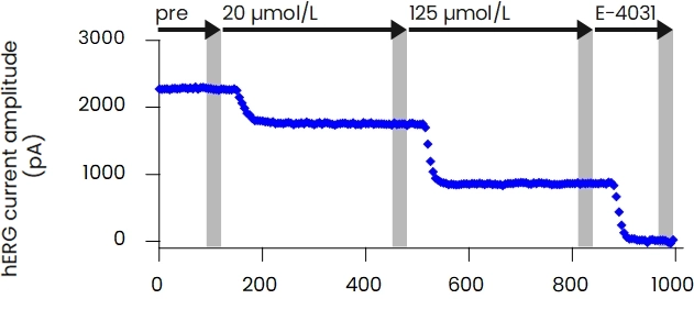
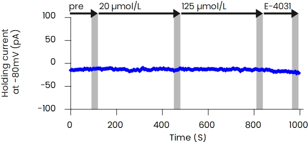
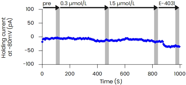
[ Representative hERG current waveforms and time plots of measurement parameters ]

Moxifloxacin Representative hERG current waveforms and time plots of measurement parameters

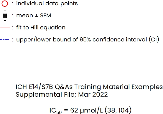
[ Concentration-response relationship ]

Moxifloxacin Concentration-response relationship
Background data for Ondansetron and Dofetilide
Ondansetron

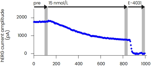
[ Representative hERG current waveforms and time plots of measurement parameters ]

Ondansetron Representative hERG current waveforms and time plots of measurement parameters

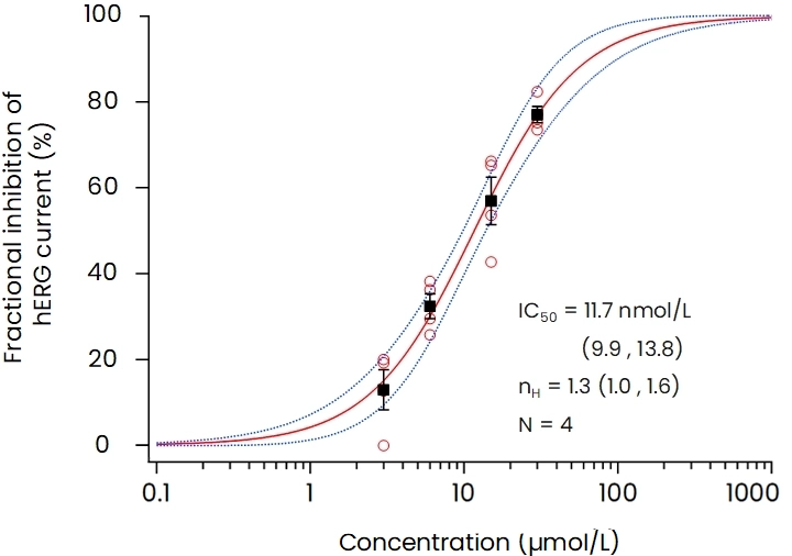
[ Concentration-response relationship ]

Ondansetron Concentration-response relationship
Dofetilide

[ Representative hERG current waveforms and time plots of measurement parameters ]

Dofetilide Representative hERG current waveforms and time plots of measurement parameters

[ Concentration-response relationship ]

Dofetilide Concentration-response relationship
Mediford’s pooled safety margin of 3 reference drugs
hERG Safety Margin and Pooled Safety Margin
Defined by Reference Drugs
ICH S7B Q&As
Training Materials
Reference Drugs Critical Concentration
(ng/mL)
Protein Binding (%) Mol wt
(g/mol)
IC50 Distribution
(µM)
Safety Margin
Moxifloxacin 1866 (1591, 2188) 40 (37, 43) 401 62 (38, 104)
N=10
23x (13, 39)
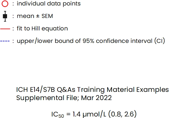
Ondansetron 249 (152, 412) 73 (71, 76) 293 1.4 (0.8, 2.6)
N=4
10x (4, 27)
Dofetilide 0.37 (0.24, 0.55) 64 (62, 66) 442 0.01 (< 0.01, 0.02)
N=4
44x (16, 117)
Pooled Safety Margin for Reference Drugs 22x (9, 51)
Threshold >51x
hERG Safety Margin and Pooled Safety Margin
Defined by Reference Drugs
Mediford Corporation
Reference Drugs Critical Concentration
(ng/mL)
Protein Binding (%) Mol wt
(g/mol)
IC50 Distribution
(µM)
Safety Margin
Moxifloxacin 1866 (1591, 2188) 40 (37, 43) 401 69.6 (63.0, 76.8)
N=1
25x (20, 30)
Ondansetron 249 (152, 412) 73 (71, 76) 293 1.10 (1.04, 1.17)
N=1
5x (3, 8)
Dofetilide 0.37 (0.24, 0.55) 64 (62, 66) 442 0.0117 (0.0099, 0.0138)
N=1
39x (25, 60)
Pooled Safety Margin for Reference Drugs 17x (4, 68)
Threshold >68x

Note: Above table is as of May 2024.

Reference:
Derek Leishman et al. Supporting an integrated QTc risk assessment using the hERG margin distributions for three positive control agents derived from multiple laboratories and on multiple occasions, Journal of Pharmacological and Toxicological Methods, available online, June 6, 2024

Multiple ion channels assay

It is regarded as a significant issue that even a drug with a useful and promising efficacy may be subject to development attrition due to the “hERG-centric” strategy in drug discovery development. Both Nav1.5 and Cav1.2 inhibition are known to reduce the risk of lethal polymorphic tachycardia (TdP) by counterbalancing the effects of hERG inhibition. Thus, evaluating hERG inhibition alone is insufficient to predict the potential occurrence of TdP associated with QT prolongation on ECG (Figure 2), and evaluating Nav1.5 and Cav1.2 inhibition has been drawing more attention. In addition, Nav1.5 and Cav1.2 data are expected to be utilized for proarrhythmic risk prediction using in silico models. Mediford is working on establishing Nav1.5 and Cav1.2 Best Practice study services under the ICH S7B Guideline Q&As 2.1 and related documents (Training Materials and FDA’s Best Practice guidance).

Figure 2:QT interval prolongation associated with hERG block

QT interval prolongation associated with hERG block
  • QT interval: Time from the start of the Q wave to the end of the T wave. QT interval prolongation is attributed to delayed repolarization (Phase 3) and prolonged plateau (Phase 2).
  • Polymorphic ventricular tachycardiaArrhythmia induced by consecutive ectopic impulses within the ventricles. Torsades de Pointes (TdP) is a form of polymorphic ventricular tachycardia in the setting manifested by QT interval prolongation.

Review the poster (2021)

Strategies for assessment using patch-clamp method under the S7B Guideline

Strategies for assessment using patch-clamp method under the S7B Guideline
Risk of QTc
prolongation
hERG assay In vivo QT
assay
Supplemental
patch-clamp assays
Other assays
Low Negative Negative
Intermediate Negative Positive
  • hERG-blocking metabolites
  • hERG trafficking inhibition

CiPA

  • Nav/Cav channel block
  • hERG trafficking inhibition (quantitative assay such as WB)

CiPA

  • hiPS-CM repolarization assay
  • In silico assessment of proarrhythmia
Intermediate Positive Negative
  • Nav/Cav channel block
  • hERG drug block kinetics using the CiPA dynamic protocol
  • hiPS-CM repolarization assay
High Positive Positive

CiPA

  • Nav/Cav channel block

CiPA

  • hiPS-CM repolarization assay
  • In silico assessment of proarrhythmia

Proarrhythmia Risk Prediction Using Human Induced Pluripotent Stem Cell-derived Cardiomyocytes

At mediford, we also offer functional and drug efficacy assay services using a multi-electrode array (MEA) system to record extracellular potentials on human iPS cell-derived cardiomyocytes (hiPS-CM). Proarrhythmic risk of test substances is assessed using the prolongation of corrected field potential duration (cFPD), which corresponds to the QT interval of the human electrocardiogram (ECG), and the occurrence of early afterdepolarization (EAD), a surrogate marker for Torsades de Pointes (TdP). This method can also be utilized as a follow-up study to assess the proarrhythmic predictivity based on multiple ion channels assays. The new ICH S7B Guideline Q&As also describe Best Practice for assessing proarrhythmic risk using hiPS-CM towards investigational new drug (IND) application.

Simultaneous data acquisition from multiple culture

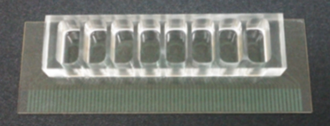
Extracellular field potential data from 8-well plate
(8 electrodes/well).

Simultaneous data acquisition from multiple culture
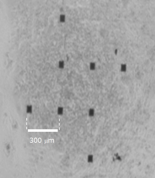
Light-microscopic image of hiPS-CM plated and electrodes
(high cell-density culture).

Automated data analysis

Comprehensive analyses
(FPD measuring, EAD detection)

Field potential waveform (before)
Emergence of EAD-like event (after 30 nM E-4031)